Describe Anthony's Capsule Stain When Would You Use It
The capsule stain employs an acidic stain and a basic stain to detect capsule production. GRAM POSITIVE bacteria and GRAM NEGATIVE bacteria.

Phone Lanyard Black Iphone Lanyard Diy Lanyard Neck Strap
Streptococcus pneumoniae Neisseria meningitidis Haemophilus influenzae Klebsiella pneumoniae are common capsulated bacteria.

. Principle of Spore Stain. A capsule is a gelatinous outer layer that is secreted by the cell and that surrounds and adheres to the cell wall. This one is another most commonly used method of Capsule staining.
Capsules may be polysaccharide glycoproteins or polypeptides depedning on the organism. Flood the smear with CRYSTAL VIOLET 1 minute then wash with water. Why dont you want to heat fix in capsule stain.
The Capsule of the bacterial cell appears as a clear halo around the stained bacterial cell body and the Dark background color depends upon the dye used India ink or Nigrosin. This is a DIFFERENTIAL STAIN. In the Anthonys Capsule stain the basic dye Crystal Violet is used to stain the background and the cell allowing the clear capsule to be observed Differential Stain This is a type of staining procedure used to distinguish one group of bacteria from another by taking advantage of the fact that certain bacteria have distinctly.
The positive capsule staining method Anthony Method uses two reagents to stain the capsular material. CAPSULE STAIN Antonys MethodPrinciple. Blot dry and find the specimen under low power.
Capsules are formed by organisms such as Klebsiella pneumoniae. The acid pH of the mordant changes the Congo red to a blue color. 100 1 rating Many bacterial secret a viscid material around the cell wall it is attached to cell layer when this organized into a well defined structure it is known as CAPSULE.
Eventually observe using oil immersion microscopy Gilmores 2nd law of microscopy. DrWhitneyHolden goes over bacterial capsules - what a capsule is and two different methods for how to stain it. When we heat fix its going to lose some moisture due to evaporation and its gonna get smaller.
Giemsa stain is a Romanowsky stain. It requires a PRIMARY STAIN and a COUNTERSTAIN. After the primary stain is applied and the smear is heated both the vegetative cell and spore will appear green.
Both the cell and the capsule become stained by the crystal violet. We review their content and use your feedback to keep the quality high. Negative staining with India ink or nigrosine is used to stain the background leaving a clear area of the cell and the capsule Counterstaining can be used to stain the cell while leaving the capsule clear.
It is not common to all organisms. For further penetration the application of heat is required. The primary stain Crystal violet is applied over a non heat fixed bacterial smear so that both the bacterial cells and capsular material take up the color of the primary stain.
We could shrink the cell. The capsule or glycocalyx is a gelatinous outer layer that is secreted by the microbe and remains stuck to it. The method involves the use of a primary stain ie 1 aqueous crystal violet.
Do not use water. Stain with crystal violet for one minute. Smear the sample at the center of a clean microscope slide.
Another method used for capsular staining is Anthony staining method devised by EE. Anthonys method uses Crystal violet 7min then rinses with 20CuSO4. Capsules stick well to glass and heat may destroy the capsule.
The capsule mordant contains a stain called acid fuchsin not the same as cabolfuschin but the same color. The basic procedure goes like this. Cells that have a heavy capsule are generally virulent and capable of producing disease since the structure protects.
It divides most of the EUBACTERIA into two large groups. Used to distinguish cells with capsules from those without. It is widely used in the microbiology laboratory for the staining of.
The capsule differs from the slime layer that most bacterial cells produce in that it is a thick detectable discrete layer outside the. Tapdistilled water decolorizing agent Safranin 25 - This is the counter stain and can be easily prepared using 25 grams of safranin O and 100ml of 95 ethanol. The capsule stain employs an acidic stain and a basic stain to detect capsule production.
Capsules appear clear or as halos if present. Allow the slide to dry air dry and then heat fix the smear. The only explanation I have found for this method states that the crystal violet stains the cell and the capsule purple and the CUSO4 decolorizes and counterstains the capsule light blue.
Wash off the excess stain with copper sulfate solution 20. Unlike most vegetative cell types that stain by common procedures the free spore because of its impervious coats will not accept the primary stain easily. Most capsules are composed of polysaccharides but some are composed of polypeptides.
I also have vi. It helps to demonstrate the presence of capsule in bacteria or yeasts. Take a heat fixed bacterial smear.
Principle of Capsule Staining Capsules stain very poorly with reagents used in simple staining and a capsule stain can be depending on the method a misnomer because the capsule may or may not be stained. Both the capsular material and the cell wall will take the colour of the stain and will appear dark blue. The acid fuschin stains the bacterial cells a pink color.
In this technique the crystal violet stain is used as the primary stain. Capsule Stain- Principle Procedure and Result Interpretation. If it shrinks your specimen will suddenly have a halo but its not a real halo.
June 5 2021 by Sagar Aryal.

Pin On Personal Care

Painting For The Home Pinterest Abstract Art Abstract Photography And Landscapes

Pin On Summer House

Raspberry Ketone Garcinia Cambogia Green Coffee Bean Tea Acai Berry Complex Raspberry Ketones Acai Berry Green Coffee

The Minimum Inhibitory Concentrations Mics Of The Extract And The Download Scientific Diagram

A Diy Turmeric Mask Is Great For Glowing Skin Acne Rosacea Eczema And Dark Circles Give It A Try Acne Skin Turmeric Mask Diy Hair Mask

Pdf Electron Microscopy Of Fish Gill Ultra Structure With Reference To Water Pollution By Municipal Wastes

Revolution Pro Colour Focus Palette Truth Or Dare 15g Intense Colors Classic Shades Top Beauty Products

Pin On Perryhill Rustics Personalized Gifts Keepsakes

Pin On Classic Game Show Hall Of Fame

Jmbfs 929 Edward Journal Of Microbiology Biotechnology And Food Sciences

Pdf Articular Cartilage Thickness Of The Humeral Head An Anatomic Study

Personalized Kids Money Box Grandchild Keepsake Gift New Etsy Kids Money Box Baby Memory Box Personalised Keepsakes

Dettol Laundry Cleanser Lavender Cleanser Antibacterial Cleaning

Pdf Oil Palm Phenolics Inhibit The In Vitro Aggregation Of B Amyloid Peptide Into Oligomeric Complexes
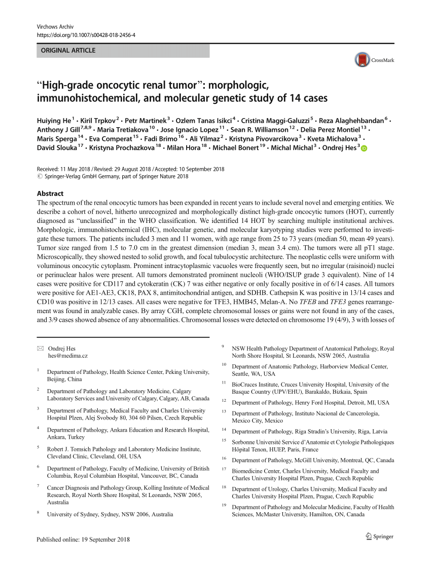
Pdf High Grade Oncocytic Renal Tumor Morphologic Immunohistochemical And Molecular Genetic Study Of 14 Cases
Pdf Recurrent Knee Pain In An Athletic Adult Multiple Schwannomas Secondary To Schwannomatosis A Case Report

Pin On Perryhill Rustics Personalized Gifts Keepsakes

Jmbfs 929 Edward Journal Of Microbiology Biotechnology And Food Sciences


Comments
Post a Comment